Retina
When it comes to your vision, there are a lot of essential components that help you see. One of these is the retina.
What is the Retina?
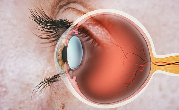
The retina is a thin layer of tissue at the back of the eye. It lines the inner surface of the eye and contains cells that capture light and transform it into signals your brain can understand. The retina works in a way that’s similar to film in a camera.
When light enters your eye through the cornea and lens, it comes to the retina. The retina then processes the light as visual information sent to the brain, allowing you to see the world.
How Does the Retina Work and Function?
The retina works because it’s made up of several types of cells. However, two types of cells are primarily essential for vision and being able to see.
These are photoreceptor cells and ganglion cells. Photoreceptor cells, called rods and cones, are what capture light. Rods are extremely sensitive to light, making it possible to see in dim lighting.
Cones allow you to see color vision and make seeing in bright lighting possible. After the photoreceptor cells capture light, it transmits the information to the ganglion cells.
The ganglion cells organize the signals from the photoreceptors before sending them through the optic nerve to the brain to be processed as images. The retina makes it possible to perceive and understand a broad range of visual details, colors, and depths. Without it, your vision would not be as rich or nuanced.
When is a Vitrectomy Necessary?
If you’re experiencing issues with your retina, you may need a vitrectomy. A vitrectomy is one of the most common retina procedures performed at Inland Valley Surgery Center.
You may need a vitrectomy if you have one of these conditions:
What to Expect During a Vitrectomy
During your vitrectomy at Inland Valley Surgery Center, you will first receive anesthesia to ensure you are comfortable during the procedure. Next, your surgeon will create small incisions in your eye to gain access to the vitreous.
After accessing the vitreous, your surgeon will use specialized instruments to remove the vitreous gel from your eye. Once the vitreous gel has been removed, your surgeon will replace it with a clear fluid to help the eye keep its shape and pressure.
The fluid may be a saline solution or a gas bubble. If a gas bubble is placed in your eye, it will absorb over time. After the vitrectomy, you may need to keep your head in a particular position for some time, especially if your surgeon used a gas bubble.
Doing so will keep the gas bubble pressed against the retinal tear or hole, allowing healing to begin. You’ll need prescription eye drops from your ophthalmologist to prevent infection and reduce inflammation while recovering.